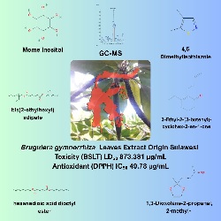
GC-MS and ADMET Profiling of Bruguiera gymnorrhiza Mangrove Leaf Extract Origin Sulawesi with Antioxidant Properties
Abstract
Keywords
Full Text:
PDFReferences
[1] T. Asaeda and A. Barnuevo, “Oxidative stress as an indicator of niche-width preference of mangrove Rhizophora stylosa,” For Ecol Manage, vol. 432, pp. 73–82, Jan. 2019, doi: 10.1016/j.foreco.2018.09.015.
[2] S. K. Das, J. K. Patra, and H. Thatoi, “Antioxidative response to abiotic and biotic stresses in mangrove plants: A review,” Int Rev Hydrobiol, vol. 101, no. 1–2, pp. 3–19, Apr. 2016, doi: 10.1002/iroh.201401744.
[3] X. Li et al., “Evaluating the physiological and biochemical responses of different mangrove species to upwelling,” Front Mar Sci, vol. 9, p. 989055, Aug. 2022, doi: 10.3389/fmars.2022.989055.
[4] N. B. Sadeer, G. Zengin, and M. F. Mahomoodally, “Biotechnological applications of mangrove plants and their isolated compounds in medicine-a mechanistic overview,” Crit Rev Biotechnol, vol. 43, no. 3, pp. 393–414, 2023, doi: 10.1080/07388551.2022.2033682.
[5] T. Sur et al., “Antioxidant and hepatoprotective properties of Indian Sunderban mangrove Bruguiera gymnorrhiza L. leaves,” J Basic Clin Pharm, vol. 7, no. 3, pp. 75–79, 2016, doi: 10.4103/0976-0105.183262.
[6] M. Haq et al., “Total phenolic contents, antioxidant and antimicrobial activities of Bruguiera gymnorrhiza,” J Med Plants Res, vol. 5, no. 17, pp. 4112–4118, 2011, doi: 10.5897/JMPR.9001250.
[7] R. Rozirwan et al., “Antioxidant activity, total phenolic, phytochemical content, and HPLC profile of selected mangrove species from Tanjung Api-Api Port Area, South Sumatra, Indonesia,” Trop J Nat Prod Res, vol. 7, no. 7, pp. 3482–3489, 2023, doi: 10.26538/tjnpr/v7i7.29.
[8] P. H. Riyadi et al., “Chemical profiles and antioxidant properties of Bruguiera gymnorrhiza fruit extracts from central sulawesi, indonesia,” Food Res, vol. 5, no. Suppl. 3, pp. 37–47, 2021, doi: 10.26656/fr.2017.5(S3).007.
[9] T. Takemura, N. Hanagata, K. Sugihara, S. Baba, I. Karube, dan Z. Dubinsky, “Physiological and biochemical responses to salt stress in the mangrove, Bruguiera gymnorrhiza,” Aquatic Botany, vol. 68, no. 1, pp. 15–28, 2000.
[10] A. Mursalim et al., “Mangrove area and vegetation condition resulting from the planting of mangroves in the Wallacea Region, Bone Bay, South Sulawesi,” IOP Conf Ser: Earth Environ Sci, vol 473, p. 012055, Mar. 2020, doi: 10.1088/1755-1315/473/1/012055.
[11] R. Hamilton et al., “A 16,000-year record of climate, vegetation and fire from Wallacean lowland tropical forests,” Quat Sci Rev, vol. 224, p. 105929, Nov. 2019, doi: 10.1016/j.quascirev.2019.105929.
[12] D. M. Ceccon et al., “Metataxonomic and metagenomic analysis of mangrove microbiomes reveals community patterns driven by salinity and pH gradients in Paranaguá Bay, Brazil,” Sci Total Environ, vol. 694, p. 133609, Dec. 2019, doi: 10.1016/j.scitotenv.2019.133609.
[13] S. Rahim et al., “Environmental quality and carrying capacity for restoration of estuarine mangrove ecosystem in the coral triangle ecoregion, Southeast Sulawesi, Indonesia,” Int J Environ Stud, vol. 81, no. 2, pp. 587–606, Mar. 2024, doi: 10.1080/00207233.2024.2326392.
[14] A. Malik, O. Mertz, and R. Fensholt, “Mangrove forest decline: consequences for livelihoods and environment in South Sulawesi,” Reg Environ Change, vol. 17, no. 1, pp. 157–169, Jan. 2017, doi: 10.1007/s10113-016-0989-0.
[15] K. Analuddin et al., “Mangrove vulnerability and blue carbon storage in the Coral Triangle Areas, Southeast Sulawesi, Indonesia,” Front Ecol Evol, vol. 12, p. 1420827, Nov. 2024, doi: 10.3389/fevo.2024.1420827.
[16] M. J. Wu, B. Xu, and Y. W. Guo, “Unusual secondary metabolites from the mangrove ecosystems: Structures, bioactivities, chemical, and bio-syntheses,” Mar Drugs, vol. 20, no. 8, p. 535, Aug. 2022, doi: 10.3390/md20080535.
[17] Y. R. Noor, M. Khazali, and I. N. N. Suryadiputra, Introduction to Mangroves in Indonesia, Kedua. Bogor: Wetlands International Indonesia Programme, 2006.
[18] D. K. Dewanto et al., “GC-MS profile of Rhizophora apiculata leaf extract from the coast of Tomini bay, Central Sulawesi with antibacterial and antioxidant activity,” Jurnal Kelautan: Indonesian Journal of Marine Science and Technology, vol. 14, no. 1, pp. 30–42, 2021, doi: 10.21107/jk.v14i1.8904.
[19] W. A. Tanod et al., “Screening of NO inhibitor release activity from soft coral extracts origin Palu Bay, Central Sulawesi, Indonesia,” Antiinflamm Antiallergy Agents Med Chem, vol. 18, no. 2, pp. 126–141, Feb. 2019, doi: 10.2174/1871523018666190222115034.
[20] L. Fu et al., “ADMETlab 3.0: an updated comprehensive online ADMET prediction platform enhanced with broader coverage, improved performance, API functionality and decision support,” Nucleic Acids Res, vol. 52, no. W1, pp. W422–W431, Jul. 2024, doi: 10.1093/nar/gkae236.
[21] P. Banerjee et al., “ProTox 3.0: a webserver for the prediction of toxicity of chemicals,” Nucleic Acids Res, vol. 52, pp. W513–W520, Jul. 2024, doi: 10.1093/nar/gkae303.
[22] Mahmiah, G. W. Sudjarwo, and A. N. Widya, “Anti-cancer potential in methanol extract of black bakau leaves (Rhizophora mucronata Poiret) using the brine shrimp lethality test (BSLT) method,” Interciencia J, vol. 45, no. 12, pp. 57–64, 2020. Accessed: Mar. 22, 2025.
[23] J. Rohmah, “Antioxidant activities using DPPH, FIC, FRAP, and ABTS methods from ethanolic extract of lempuyang gajah rhizome (Zingiber zerumbet (L.) Roscoeex Sm.),” Jurnal Kimia Riset, vol. 7, no. 2, pp. 152–166, 2022, doi: 10.20473/jkr.v7i2.34493.
[24] S. A. Sami, “New insights into the identification of bioactive compounds from Willughbeia edulis Roxb. through GC–MS analysis,” Beni Suef Univ J Basic Appl Sci, vol. 11, pp. 8–11, 2022, doi: 10.1186/s43088-022-00270-8.
[25] H. M. El-Naggar, A. M. Shehata, and M. A. A. Morsi, “Micropropagation and GC–MS analysis of bioactive compounds in bulbs and callus of white squill,” In Vitro Cell Dev Biol-Plant, vol. 59, no. 1, pp. 154–166, 2023, doi: 10.1007/s11627-023-10333-9.
[26] A. I. Prihantini et al., “Phytochemical test and antibacterial activity of pranawija (Euchresta horsfieldii (Lesch.) Benn.),” Jurnal Ilmu Kehutanan, vol. 12, pp. 223–233, 2018, doi: 10.22146/jik.40157.
[27] Usman et al., “Antidiabetic activity of leaf extract from three types of mangrove originating from Sambera coastal region Indonesia,” Res J Pharm Technol, vol. 12, no. 4, pp. 1707–1712, 2019, doi: 10.5958/0974-360X.2019.00284.1.
[28] P. Revathi, T. Jeyaseelansenthinath, and P. Thirumalaikolundhusubramaian, “Preliminary phytochemical screening and GC-MS analysis of ethanolic extract of mangrove plant-Bruguiera cylindrica (Rhizho) L,” Int J Pharm Phyto Res, vol. 6, no. 4, pp. 729–740, 2014.
[29] K. Arora et al., “Mangroves: A novel gregarious phytomedicine for diabetes,” IJRDPL, vol. 3, no. 6, pp. 1244–1257, 2014. Accessed: Feb. 18, 2025.
[30] W. M. Bandaranayake, “Bioactivities, bioactive compounds and chemical constituents of mangrove plants,” Wetl Ecol Manag, vol. 10, no. 6, pp. 421–452, 2002, doi: 10.1023/A:1021397624349.
[31] H. Ashihara et al., “Pyridine salvage and nicotinic acid conjugate synthesis in leaves of mangrove species,” Phytochem, vol. 71, no. 1, pp. 47–53, Jan. 2010, doi: 10.1016/j.phytochem.2009.09.033.
[32] Rozirwan et al., “Phytochemical profile and toxicity of extracts from the leaf of Avicennia marina (Forssk.) Vierh. collected in mangrove areas affected by port activities,” S Afr J Bot, vol. 150, pp. 903–919, Nov. 2022, doi: 10.1016/j.sajb.2022.08.037.
[33] K. Kalasuba et al., “Red mangrove (Rhizophora stylosa Griff.)—A review of its botany, phytochemistry, pharmacological activities, and prospects,” Plants, vol. 12, no. 11, p. 2196, Jun. 2023, doi: 10.3390/plants12112196.
[34] Rozirwan et al., “Insecticidal activity and phytochemical profiles of Avicennia marina and Excoecaria agallocha leaves extracts,” ILMU KELAUTAN: Indonesian Journal of Marine Sciences, vol. 28, no. 2, pp. 148–160, Jun. 2023, doi: 10.14710/ik.ijms.28.2.148-160.
[35] N. M. Alikunhi, R. Narayanasamy, and K. Kandasamy, “Fatty acids in an estuarine mangrove ecosystem,” Rev Biol Trop, vol. 58, no. 2, pp. 577–587, 2010, doi: 10.15517/rbt.v58i2.5230.
[36] X. Zhang et al., “Vomifoliol isolated from mangrove plant Ceriops tagal inhibits the NFAT signaling pathway with CN as the target enzyme in vitro,” Bioorg Med Chem Lett, vol. 48, p. 128235, Sep. 2021, doi: 10.1016/j.bmcl.2021.128235.
[37] G. Eswaraiah et al., “Identification of bioactive compounds in leaf extract of Avicennia alba by GC-MS analysis and evaluation of its in-vitro anticancer potential against MCF7 and HeLa cell lines,” J King Saud Univ Sci, vol. 32, no. 1, pp. 740–744, Jan. 2020, doi: 10.1016/j.jksus.2018.12.010.
[38] T. Sivakumar, “Phytochemical screening and gas chromatography-mass spectroscopy analysis of bioactive compounds and biosynthesis of silver nanoparticles using sprout extracts of Vigna radiata L. and their antioxidant and antibacterial activity,” Asian J Pharm Clin Res, vol. 12, no. 2, pp. 180–184, 2019, doi: 10.22159/ajpcr.2019.v12i2.29253.
[39] A. Sunita and S. Manju, “Phytochemical examination and GC-MS analysis of methanol and ethyl- acetate extract of root and stem of Gisekia pharnaceoides Linn. (Molluginaceae) from Thar Desert, Rajasthan, India,” Res J Pharm Biol Chem Sci, vol. 8, no. 4, pp. 168–174, 2017. Accessed: Feb. 19, 2025.
[40] R. B. Venkata et al., “Antibacterial, antioxidant activity and GC-MS analysis of Eupatorium odoratum,” Asian J Pharm Clin Res, vol. 5, no. Suppl 2, pp. 99–106, 2012. Accessed: Feb. 19, 2025. [Online]. Available: https://www.innovareacademics.in/journal/ajpcr/Vol5Suppl2/940.pdf.
[41] S. Mitra et al., “A study on phytochemical profiling of Avicennia marina mangrove leaves collected from Indian Sundarbans,” Sci Total Environ, vol. 4, p. 100041, Dec. 2023, doi: 10.1016/j.scenv.2023.100041.
[42] IARC Working Group, “Di(2-ethylhexyl) adipate,”. Accessed: Oct. 28, 2024.
[43] S. K. Maiti and A. Chowdhury, “Effects of anthropogenic pollution on mangrove biodiversity: A review,” J Environ Prot (Irvine, Calif), vol. 4, no. 12, pp. 1428–1434, 2013, doi: 10.4236/jep.2013.412163.
[44] Y. Chen et al., “Di-2-ethylhexyl phthalate (DEHP) degradation and microbial community change in mangrove rhizosphere gradients,” Sci Total Environ, vol. 871, p. 162022, May 2023,
doi: 10.1016/j.scitotenv.2023.162022.
[45] A. Talukdar et al., “Microplastics in mangroves with special reference to Asia: Occurrence, distribution, bioaccumulation and remediation options,” Sci Total Environ, vol. 904, p. 166165, Dec. 2023, doi: 10.1016/j.scitotenv.2023.166165.
[46] H. Moniuszko et al., “Accumulation of plastics and trace elements in the mangrove forests of Bima City Bay, Indonesia,” Plants, vol. 12, no. 3, p. 462, Feb. 2023, doi: 10.3390/plants12030462.
[47] A. L. da S. Pontes et al., “Phthalates in Avicennia schaueriana, a mangrove species, in the State Biological Reserve, Guaratiba, RJ, Brazil,” Env Adv, vol. 2, p. 100015, Dec. 2020, doi: 10.1016/j.envadv.2020.100015.
[48] I. N. Vikhareva, G. K. Aminova, and A. K. Mazitova, “Ecotoxicity of the adipate plasticizers: Influence of the structure of the alcohol substituent,” Moeculesl, vol. 26, no. 16, p. 4833, Aug. 2021, doi: 10.3390/molecules26164833.
[49] A. V. Kletskov et al., “Isothiazoles in the design and synthesis of biologically active substances and ligands for metal complexes,” Synthesis, vol. 52, no. 2, pp. 159–188, 2020,
doi: 10.1055/s-0039-1690688.
[50] M. Haroun et al., “Discovery of 5-methylthiazole-thiazolidinone conjugates as potential anti-inflammatory agents: Molecular target identification and in silico studies,” Molecules, vol. 27, no. 23, p. 8137, Dec. 2022,
doi: 10.3390/molecules27238137.
[51] Z. Zhang et al., “Design, synthesis, and SAR study of novel 4,5-dihydropyrazole-thiazole derivatives with anti-inflammatory activities for the treatment of sepsis,” Eur J Med Chem, vol. 225, p. 113743, Dec. 2021, doi: 10.1016/j.ejmech.2021.113743.
[52] S. Y. Shin et al., “Anticancer activities of cyclohexenone derivatives,” Appl Biol Chem, vol. 63, no. 1, p. 82, Dec. 2020, doi: 10.1186/s13765-020-00567-1.
[53] Y. Wang et al., “A novel compound C12 inhibits inflammatory cytokine production and protects from inflammatory injury in vivo,” PLoS One, vol. 6, no. 9, p. e24377, Sep. 2011, doi: 10.1371/journal.pone.0024377.
[54] H. B. Küçük et al., “Synthesis and biological activity of new 1,3-dioxolanes as potential antibacterial and antifungal compounds,” Molecules, vol. 16, no. 8, pp. 6806–6815, Aug. 2011, doi: 10.3390/molecules16086806.
[55] P. Chawla, R. Singh, and S. K. Saraf, “Effect of chloro and fluoro groups on the antimicrobial activity of 2,5-disubstituted 4-thiazolidinones: A comparative study,” Med Chem Res, vol. 21, no. 10, pp. 3263–3271, Oct. 2012, doi: 10.1007/s00044-011-9864-1.
[56] Y. Hai et al., “The intriguing chemistry and biology of sulfur-containing natural products from marine microorganisms (1987–2020),” Mar Life Sci Technol, vol. 3, no. 4, pp. 488–518, Nov. 2021, doi: 10.1007/s42995-021-00101-2.
[57] J. Xie et al., “Synthesis of novel 2-methyl-3-furyl sulfide flavor derivatives as efficient preservatives,” RSC Adv, vol. 11, no. 41, pp. 25639–25645, Jul. 2021, doi: 10.1039/d1ra04207f.
[58] J. Basbous et al., “Dihydropyrimidinase protects from DNA replication stress caused by cytotoxic metabolites,” Nucleic Acids Res, vol. 48, no. 4, pp. 1886–1904, Feb. 2020, doi: 10.1093/nar/gkz1162.
[59] N. Roy et al., “Different routes for the construction of biologically active diversely functionalized bicyclo[3.3.1]nonanes: an exploration of new perspectives for anticancer chemotherapeutics,” RSC Adv, vol. 13, no. 32, pp. 22389–22480, Jul. 2023, doi: 10.1039/d3ra02003g.
[60] M. Yamauchi et al., “DPPH measurements and structure—activity relationship studies on the antioxidant capacity of phenols,” Antioxidants, vol. 13, no. 3, p. 309, Mar. 2024, doi: 10.3390/antiox13030309.
[61] A. F. Al-Rubaye, M. J. Kadhim, and I. H. Hameed, “Determination of bioactive chemical composition of methanolic leaves extract of Sinapis arvensis using GC-MS technique,” IJTPR, vol. 9, no. 2, pp. 163–178, 2017, doi: 10.25258/ijtpr.v9i02.9055.
[62] B. Chunsong et al., “Exploring expected values of topological indices of random cyclodecane chains for chemical insights,” Sci Rep, vol. 14, no. 1, p. 10065, Dec. 2024, doi: 10.1038/s41598-024-60484-x.
[63] K. Surana et al., “Benzophenone: A ubiquitous scaffold in medicinal chemistry,” Med Chem Comm, vol. 9, no. 11, pp. 1803–1817, 2018, doi: 10.1039/C8MD00300A.
[64] T. Marinov, Z. Kokanova-Nedialkova, and P. T. Nedialkov, “Naturally occurring simple oxygenated benzophenones: Structural diversity, distribution, and biological properties,” Diversity, vol. 15, no. 10, p. 1030, Oct. 2023, doi: 10.3390/d15101030.
[65] S. R. M. Ibrahim et al., “Benzophenones-natural metabolites with great Hopes in drug discovery: structures, occurrence, bioactivities, and biosynthesis,” RSC Adv, vol. 13, no. 34, pp. 23472–23498, Aug. 2023, doi: 10.1039/d3ra02788k.
[66] P. Das et al., “Synthesis and biological evaluation of fluoro-substituted spiro-isoxazolines as potential anti-viral and anti-cancer agents,” RSC Adv, vol. 10, pp. 30223–30237, Aug. 2020, doi: 10.1039/d0ra06148d.
[67] L. Lingfa, A. Tirumala, and S. Ankanagari, “GC-MS profiling of anticancer and antimicrobial phytochemicals in the vegetative leaf, root, and stem of Withania somnifera (L.) Dunal,” Int J Second Metab, vol. 11, no. 1, pp. 63–77, 2023, doi: 10.21448/IJSM.1256932.
[68] A. Staudt et al., “Biocatalytic production of citronellyl esters by different acylating agents: : A kinetic comparison study,” in 7th Brazilian Conference on Natural Product/ XXXIII RESEM Proceedings, Rio de Janeiro, Brazil, Jun. 2023. Accessed: Nov. 8, 2024. .
[69] J. Dulsat et al., “Evaluation of free online ADMET tools for academic or small biotech environments,” Molecules, vol. 28, no. 2, p. 776, Jan. 2023, doi: 10.3390/molecules28020776.
[70] Z. Wang et al., “Determination of in vitro permeability of drug candidates through a Caco-2 cell monolayer by liquid chromatography/tandem mass spectrometry,” J Mass Spectrom, vol. 35, no. 1, pp. 71–76, 2000,
doi:10.1002/(SICI)1096-9888(200001)35:1<71:AID-JMS915>3.0.CO;2-5.
[71] M. Gupta, J. Feng, and G. Bhisetti, “Experimental and computational methods to assess central nervous system penetration of small molecules,” Molecules, vol. 29, no. 6, p. 1264, Mar. 2024, doi: 10.3390/molecules29061264.
[72] J. Wu et al., “Implications of plasma protein binding for pharmacokinetics and pharmacodynamics of the γ-secretase inhibitor RO4929097,” Clin Cancer Res, vol. 18, no. 7, pp. 2066–2079, Apr. 2012, doi: 10.1158/1078-0432.CCR-11-2684.
[73] P. Manikandan and S. Nagini, “Cytochrome P450 structure, function and clinical significance: A review,” Curr Drug Targets, vol. 19, no. 1, pp. 38–54, 2017, doi: 10.2174/1389450118666170125144557.
[74] C. Clarkson et al., “In vitro antiplasmodial activity of medicinal plants native to or naturalised in South Africa,” J Ethnopharmacol, vol. 92, no. 2–3, pp. 177–191, Jun. 2004, doi: 10.1016/j.jep.2004.02.011.
[75] M. Blois, “Antioxidant determinations by the use of a stable free radical,” Nature, vol. 181, no. 4617, pp. 1199–1200, 1958, doi: 10.1038/1811199a0.
[76] R. Devitria, H. Sepriyani, and S. Sari,Refbacks
- There are currently no refbacks.


